Disclaimer
All content provided on this blog is representation of the blog owner and not Franciscan University of Steubenville. The information on this site is purely used for education purposes. The owner of this blog makes no representation as to accuracy or completeness of any information on this site or found by following any link on this site. The owner will not be liable for errors or omissions in this information nor for availability of this information. The owner will not be liable for any losses, injuries, or damages from the display or use of this information.
Privacy
The owner of this blog does not share personal information with third-parties nor does the owner store information is collected about your visit for use other than to analyze content performance through the use of cookies, which you can turn off at anytime by modifying your Internet's browser's settings. The owner is not responsible for the republishing of the content found on this blog on other Web sites or media without permission.
Blog Comments
The owner of this blog reserves the right to edit or delete any comments submitted to this blog without notice due to:
1. Comments deemed to be spam or questionable spam
2. Comments including profanity
3. comments containing language or concepts that could be deemed offensive
4. Comments that attack a person individually
This policy is subject to change anytime
Ari and Johanna's Medical Microbiology Lab Report
Sunday, December 15, 2013
Friday, December 6, 2013
Last Class Sad, but Last Experiment = COOL!!!!
12/3
Today, sadly was our last lab, but we went out with a bang! We did a virus test to see if our "student" tested positive or negative for the presence of a pathogen. The first three cups, have plus signs on them, they are our "fail safe" that is so we can make sure that we will have a positive reaction (letting us know that everything works). The middle ones (with the "-" sign) those are the negative results. Now what we had to do was test two "students" to see if they were negative or positive. Student number 15 or (FN) tested negative. Student 32 tested positive. The "positive" is indicated by the blue color, and the negative is indicated by no color change at all.
After:
Today, sadly was our last lab, but we went out with a bang! We did a virus test to see if our "student" tested positive or negative for the presence of a pathogen. The first three cups, have plus signs on them, they are our "fail safe" that is so we can make sure that we will have a positive reaction (letting us know that everything works). The middle ones (with the "-" sign) those are the negative results. Now what we had to do was test two "students" to see if they were negative or positive. Student number 15 or (FN) tested negative. Student 32 tested positive. The "positive" is indicated by the blue color, and the negative is indicated by no color change at all.
The second experiment we did was a meat purity experiment. We took a agar and injected it with dye (red, green), barium chloride and potassium sulfate. The next slot we injected with Bovine albumin, Goat Anti-horse albumin, Goat Anti-bovine Albumin, Goat Anti-swine Albumin. The last one we injected with Hamburger Extract, Goat Anti-Horse Albumin, Goat Anti-bovine Albumin, Goat Anti-swine Albumin. Now what these were supposed to do is if you put them into a meat they were "anti" or against, then there would form an arch between the two "fighting." However, our materials were old and that is why we suspect that ours didn't work. Here is a picture of it none the less. You can see in the colored one, the black portion between the colors is what the rest should have looked like had they been working.
Before:
After
-JTA and ART
Sunday, December 1, 2013
UV light, Drugs, Anaerobic Chamber, Yogurt, Retesting and BACTERIA REVEALING!!!! :D
UV light can kill the bacteria, here is a picture of me using the UV light to kill the bacteria.
DRUGS:
Novobiocin - it did not work it is a nucleic acid synthesis inhibitor
Erythromycin - nothing happened, this is translation ribosome inhibitor
Aminoglycoside - (this particular antibiotic binds the 30s ribosome and allows it to misread the RNA) this seemed to work best on all gram negative bacteria, it worked on ours 2cm
Tetracycline - this breaks the attachment of tRNA, this worked well it was 4cm
Penicillin - this breaks down the cell wall. This worked very well with our bacteria, it was 4cm
Cefoxtin - this also breaks the cell wall, and it did a good job on ours it was 4cm too.
Anaerobic Chamber:
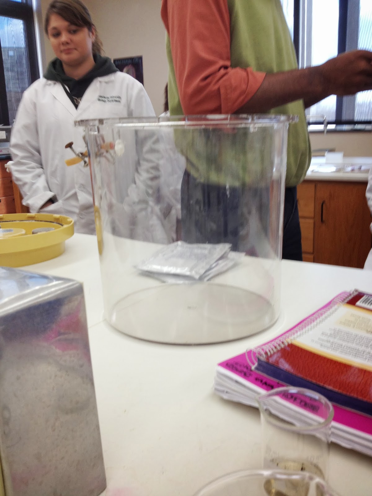
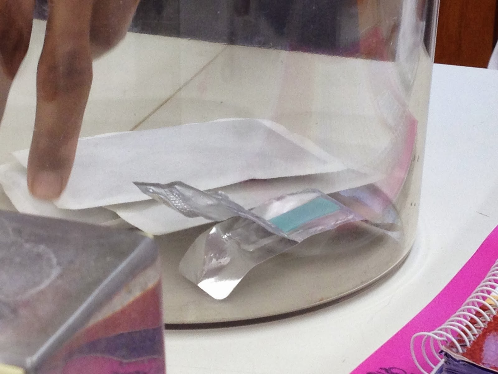
The blue strip of paper indicates that oxygen is present. It was white previously indicating that no oxygen was present. What we did in this experiment was suck the oxygen out of the chamber. This would test if our bacteria could grow in side. Our bacteria hardly grew, meaning it does need oxygen, but it can do without it just it will not thrive.
Yogurt:
We had previously mixed some yogurt into cold milk and then into milk that had been heated. Our result was that there was yogurt produced in both, but the milk that had been heated was less sour than the one that had not been. Maybe this was because the temperature killed the bacteria that secrete the sourness as a taste? We are not really sure.
RETESTING
We retested our bacteria for the Lactose test and the urease test. Our results were lactose negative, urease positive:
here is a picture of the urease because it was positive, it had a extreme change in color! This surprised us, because our last test result was negative. We assume that we must not have inoculated it or something because how else could it have produced such a different result? But any how, after following the flow chart this led us to the conclusion that our unknown bacteria was Proteus Vulgaris! This is a ear infection bacteria :)
Nose Swab
We did a nose swab today and put it on a Manitol salt plate to see if we had staph in our noses.
Ours is the one on the bottom, Ari doesn't have any staph in her nose because there is no yellow. The cool thing was that Ari had a cold. So obviously staph is not always present when you have a cold.
The second thing we did in class was put our unknown bacteria on a plate and putting various cleaning agents on them to see if they work against our bacteria.
Going from the upper left going across, it goes2% lysol: worked well the circumference was about 4cm
Spray lysol: did not work at all
Antibacterial hand soap: worked very well had 6cm
Lysol wipes: Worked pretty well was 4cm
Antiseptic mouthwash: did not work at all
Hand sanitizer: did not work at all
Clorox cleanup: Worked fairly well 2cm
Toothpaste: worked fairly well 2cm
Ari and I wanted to try again with the ones that worked very well so we retried it on some bacteria that we knew would really work.
the top is 2% lysol. It worked very well at killing the bacteria
far right is lysol wipe and it worked sort of well, but some of the bacteria started growing underneath the wipe, this must have been where the liquid cleaner must not have touched.
Bottom is is the antibacterial handsoap, and this one seemed to work the best. It ate away at the bacteria.
Results
Test 1
Blood Agar Plate:
The purpose of this test is to distinguish which bacteria possess the ability to lyse red blood cells.
Result:
Our bacteria showed no lysing of the RBC's. The official name is gamma- hemolysis.
Test 2
EMB (Eosin Methylene):
The purpose of this test is to determine if your bacteria is really gram negative. The gram negative bacteria is indicated by growth on the plate.
Result: It is hard to see, but there is a "wee" dot of growth in this picture! Our bacteria was really tiny, so maybe that was why the growth was barley there? We are not sure, but either way we are definitely positive that our bacteria is gram negative.
Test 3
Mannitol Salt Agar:
This test is used to test the presence of a staph bacteria. Our bacteria was not staphilococcus bacteria. A positive test would yield a growing yellow mass of bacteria.
Test 4
MacConkey Agar
This was to see if our bacteria ferments lactose. The test was positive. I kiddingly called our bacteria a "fat kid" because it really liked sugar :).
Test 5
PEA
This is a selective medium only gram positive bacteria grows on this plate. Our plate was positive, therefore our bacteria is most definitely a gram positive. Okay, we know it doesn't look like it, but because our bacteria is so tiny and I mean TINY, that when it does grow it seems like it hardly grew.
Test 6
Dnase
This test is to see if bacteria hydrolyzes a lipid. Our test was negative. Our bacteria does not hydrolyze
lipids.
Test 7
Thioglycolate:
This test is to see if our bacteria requires/needs oxygen. We came to the conclusion that our bacteria is facultative because it grew all over the tube.
Wednesday, October 23, 2013
Lots of Inoculations!
1. Blood Agar Plate
2. Eosin Methylene Blue Agar (EMB)
3. Mannitol Salt Agar
4. MacConkey Agar
5. Phenylethyl Alcohol AGAR (PEA)
Glucose, Lactose, Sucrose and TSI Enzyme Presence
10/15
The "Before" picture of the tubes are from left to right: Sucrose, Glucose, Lactose and TSA
All tests were to determine if our bacteria could ferment a particular carbohydrate, in this case Lactose, Sucrose and Glucose.
Glucose: Positive
It changed color to yellow indicating acid and it had a bubble indicating gas.
See the bubble?
Lactose: Positive
It changed color to yellow indicating acid and it had a bubble indicating gas.
Pretty yellow no?
Sucrose: Negative
Sadly, this test was negatory! But, at least it was still a pretty color! :) The VERY bright pink indicates no breaking down of sucrose!
TSI: Positive
To see which one, glucose or sucrose, that the bacteria really eats.
Our results were: Acid slant/alkaline but, with Hydrogen disulphide gas (Black at the bottom).
Until next time then!
JTA and ART
:)
Subscribe to:
Comments (Atom)


