Sunday, September 22, 2013

Unknown A

09/17/13

Last week, at the end of class, Dr. P gave us a test tube filled with an unknown bacteria. First we transferred some of the unknown bacteria into another test tube as our "stock" colony.
 Next we preformed a Gram stain on our sample to identify the shape and the thickness of the cell wall.


Our conclusion was that the bacteria has a gram negative cell wall, and their shape is small rods. VERY tiny! 

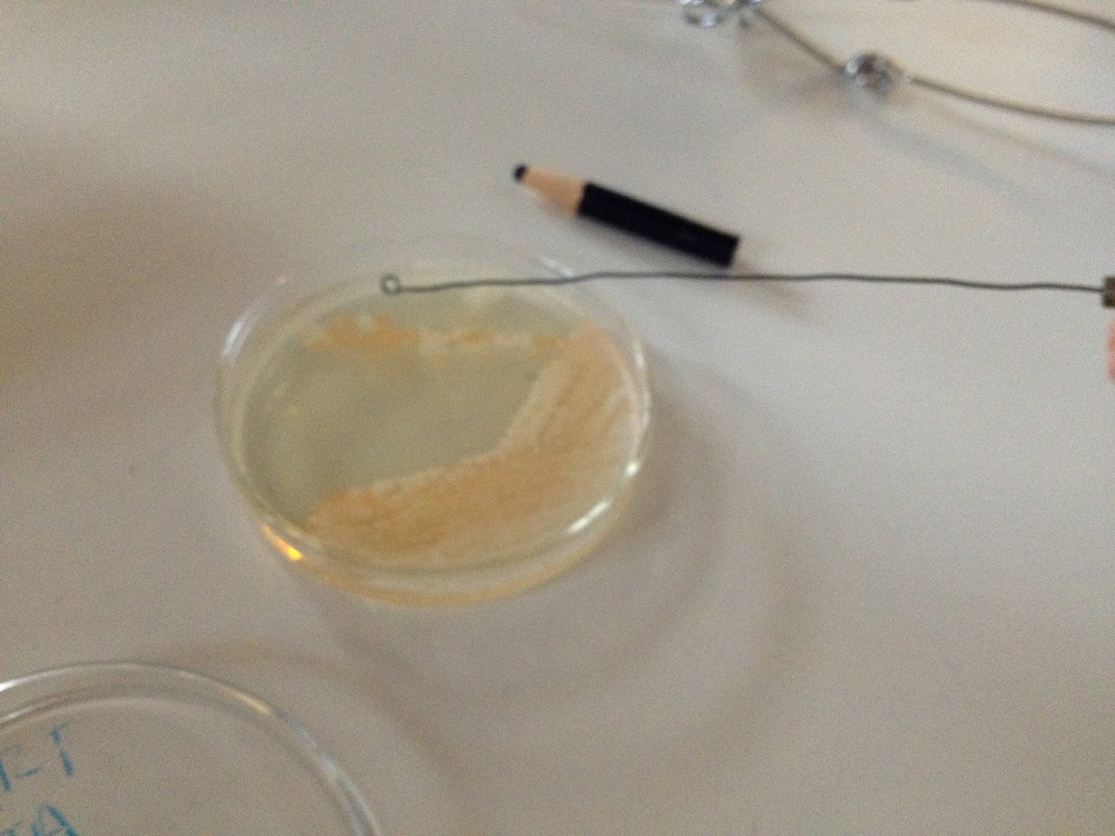
Perhaps that is why our unknown didn't seem to grow that fast after a whole weekend.

9/19/13

During this cool class, we preformed (for the first time) the negative staining. The purpose of negative staining is to see the shape of hard to see bacteria. Like ours. This is much like simple staining, the only difference is that you don't fix the bacteria to the slide. Instead, you use a very black stain to view them.

 We had to spread the stain out so it could dry... a very tricky process because we had to make sure there was a thin layer.

 This was our result, looks kind of like a night sky doesn't it ...


That is... until Dr. P fixed it! You can see the bacteria much better now! 


By the way, those are live bacteria you are looking at!

After this, our next assignment was to capsule stain the bacteria.... which totally failed! A capsule stain involves the same steps as up above, using a negative stain, but after wards using safranin to try to really distinguish the capsule. We must have rinsed off the safranin too much because this is how it turned out. You can't see much, but here is a picture anyway! 


Until later then,

JTA and ART

Sunday, September 15, 2013

Gram Staining

09/12
 
Goal: Gram staining for the purpose of determining gram negative/gram positive bacteria.

  In class today we learned how to gram stain bacteria. This similar to the simple stain, but with several extra steps of the same nature. This time after putting on the crystal violet stain we had to wash it off after only 20 seconds and put on some gram iodine.


Afterwards we rinsed the gram iodine off and applied drops of decolorizing agent until the blue stopped running. 


      After this we applied a second stain, safranin, and let that sit for one minute. We rinsed off the excess stain and prepared a slide for viewing. On viewing the slide our bacteria seemed to have a lot of clearly defined spores, and Dr. P told us that we had blown our bacteria up due to heating it up too much. Whoops! Sorry lil' bacteria! 
      This was the final result after doing everything correctly. We came to the conclusion that our cell wall was gram negative, because of the pink color. Had it been gram positive the bacteria would have been completely blue. 


JTA and ART

Sunday, September 8, 2013

Hand Washing Phenomena: Colony Isolation

09/03/13
        Background Story: Dr. P, the first day of class, handed over petri dishes and told us to stick our uncleaned thumb onto it, then, wash our hands and touch it in a different place to observe how good we were at washing our hands. What trickery was this? Of course our befores were going to be dirtier than our afters! Psych! Once we did this the petri dish was put in the incubator and after 48 hours of incubating this was the terrifying result:


Conclusion: there is something in the water or on the paper towels, because we were all dirtier after having washed out hands.

        We then used this to learn how to isolate colonies of bacteria via streak plate technique. Once the technique was preformed we put the new petri dish back in the room temperature incubator to await our isolated (hopefully!) colonies.

09/05/13
       Did the streak plate technique do its job? Were we successful in our endeavor? Look and see:

YEA! We successfully isolated a colony, and plus, it was pretty! Didn't smell so great, though!
Next line of business: learning to stain the isolated bacteria. We used the pink colony.

Since a picture is worth a thousand words, we decided to show you up close and PERSONAL!

Don't forget to sterilize that inoculating loop! :)

                                             Taking a sample of the bacteria for the slide

We smeared the bacteria from the loop onto the water on this slide. We let the slide air dry. Then we quickly ran it through a low heat flame to ensure that the bacteria were fixed on the slide.

                                           Next step dying:


                                         Then washing:

Blotting off excess water...


Now we get to look at it through the microscope to see if we did it correctly ;) WHOOPEEE!


This is what we saw at first... and then our instructor so kindly reminded us that the point of an OIL EMERSION lens (which happened to be the lens we were looking through)  is to actually use OIL! Who would have thunk? Whoops.... So after being enlightened this was the much clearer result... as our instructor said its like a person with cataracts vs. a person with 20/20 vision. He was right!


If you look closely, you can see bacteria filled with spores... oh the power of the oil emersion lens! YAH, Spores ..... gross!

Conclusion: We definitely learned a lot and were grossed out in the process. All you Bio majors out there, know why we got freaked out by spores. :) Anywho, we learned how to better our microbiology experience by proper use of lab tools.... and to where gloves EVERYWHERE from now on. And masks. *<:-)

Until Next Time Then,
JTA and ART

Collecting Specimens from the Environment

08/27/13
    DISCLAIMER: The views expressed herein are the sole opinions of the authors and do not reflect Franciscan University of Steubenville's views. 
     
    Today we went out into the wonderful world of microbes and collected specimen for a germ inquiry. We collected two samples. The first was from the bottom of Ari's shoe (which has been EVERYWHERE!!! farm, airport, beach ... you name it!), and for the other sample a knight in shining armor went into the boys bathroom for us and swabbed the stall handle.
      Once we had retrieved the samples, we wiped them on the agar solution on different petri dishes; put them in the room temperature incubator to await, with excited anticipation, the resulting growth.

08/29/13
Upon arrival in the lab, after washing our hands of course, we retrieved our petri dishes and saw immense growth! EWWWW!


Bottom of shoe                                                           Boy's Bathroom Handle

(Good job Boys! Now we want to see the girls, do a little comparison! Eh?)


We expected that the specimen collected from the boy's restroom would be the worst of the two, but were surprised to discover just the opposite. Perhaps the restroom had just been cleaned? We definitely were reminded of the fact that bacteria are ubiquitous (everywhere)!

What interesting (and/or disgusting) things shall we see next????

~ JTA, ART