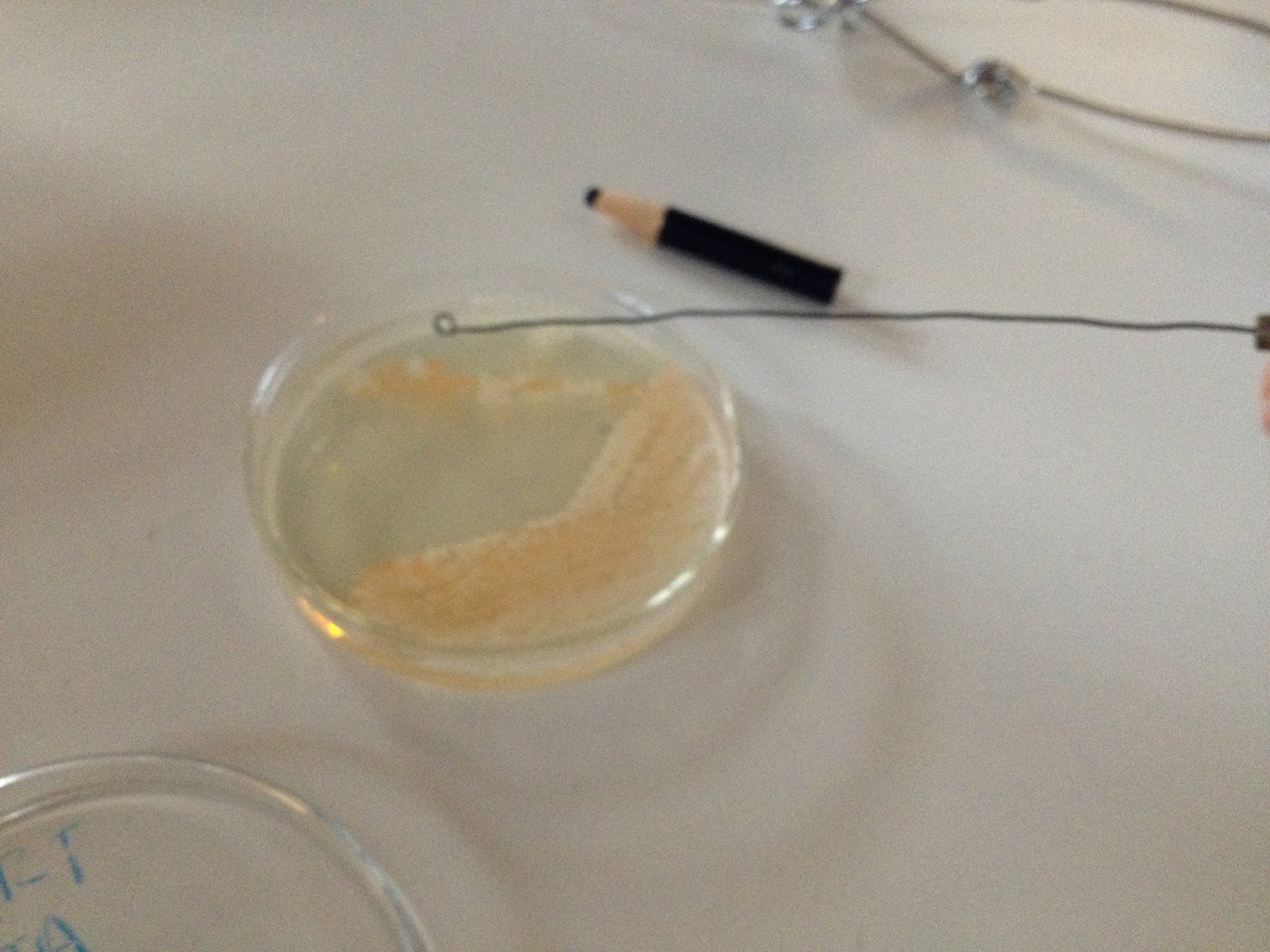
Last week, at the end of class, Dr. P gave us a test tube filled with an unknown bacteria. First we transferred some of the unknown bacteria into another test tube as our "stock" colony.
Next we preformed a Gram stain on our sample to identify the shape and the thickness of the cell wall.
Our conclusion was that the bacteria has a gram negative cell wall, and their shape is small rods. VERY tiny!
Perhaps that is why our unknown didn't seem to grow that fast after a whole weekend.
9/19/13
During this cool class, we preformed (for the first time) the negative staining. The purpose of negative staining is to see the shape of hard to see bacteria. Like ours. This is much like simple staining, the only difference is that you don't fix the bacteria to the slide. Instead, you use a very black stain to view them.
We had to spread the stain out so it could dry... a very tricky process because we had to make sure there was a thin layer.
That is... until Dr. P fixed it! You can see the bacteria much better now!
By the way, those are live bacteria you are looking at!
After this, our next assignment was to capsule stain the bacteria.... which totally failed! A capsule stain involves the same steps as up above, using a negative stain, but after wards using safranin to try to really distinguish the capsule. We must have rinsed off the safranin too much because this is how it turned out. You can't see much, but here is a picture anyway!
Until later then,
JTA and ART


